

Table of Contents
- What Is Sudden Vision Loss and Why It Matters
- Common Medical Causes of Sudden Vision Loss
- Signs of Retinal Detachment and Other Critical Conditions
- Symptoms of Ocular Stroke and Eye Stroke Warning Signs
- When to Go to the Emergency Room for Eye Pain and Vision Changes
- Diagnostic Tests and What to Expect During Emergency Evaluation
- Recovery and Long-Term Outlook After Sudden Vision Loss
- Conclusion
Last Updated: August 19, 2026
What Is Sudden Vision Loss and Why It Matters
Sudden vision loss is a dramatic, rapid change in eyesight occurring within hours or minutes, not the gradual blurring of aging or needing new glasses. At Clear Vision San Antonio, we emphasize that sudden vision loss is never something to monitor at home or wait out. The speed of onset often signals a serious underlying condition requiring urgent intervention.
Many causes of sudden vision loss can lead to permanent damage if not treated within hours. Your eye’s delicate structures, the retina, optic nerve, and blood vessels, are exquisitely sensitive to interruption in blood flow or trauma. When something goes wrong suddenly, time becomes your most valuable asset. The difference between seeking care immediately and waiting even a few hours can determine whether you retain your vision or experience lasting impairment.
This guide from Clear Vision San Antonio breaks down the medical causes, warning signs, and exact steps you should take if sudden vision loss happens to you or someone in your care.
Common Medical Causes of Sudden Vision Loss
Sudden vision loss stems from vascular events (interruption of blood flow), retinal problems, and inflammatory or infectious conditions.
Retinal artery occlusion, often called an eye stroke, occurs when a blood clot blocks blood flow to the retina. This is a medical emergency comparable to a stroke in the brain. The retina starves for oxygen within minutes, and irreversible damage begins quickly. Patients typically describe sudden, complete vision loss in one eye, sometimes preceded by amaurosis fugax, a temporary blackout lasting seconds to minutes.
Retinal detachment happens when the light-sensitive tissue at the back of your eye pulls away from its supporting layer. This can occur spontaneously, especially in people with myopia (nearsightedness) or a history of eye trauma. Early signs include floaters (spots drifting across your vision) and flashes of light, followed by a shadow or curtain effect creeping across your visual field.
Vitreous hemorrhage involves bleeding into the gel-like substance filling your eye. This can result from trauma, diabetes-related bleeding, or vitreous detachment. Patients often see sudden floaters or a shower of dark spots, sometimes accompanied by blurred vision.
Giant cell arteritis (temporal arteritis) is an inflammatory condition affecting blood vessels, particularly in older adults. It can cause sudden vision loss in one or both eyes and typically brings scalp tenderness, jaw pain while chewing, and general malaise.
Acute angle-closure glaucoma occurs when fluid pressure inside the eye spikes dramatically as the drainage angle becomes blocked. This causes severe eye pain, blurred vision, halos around lights, and sometimes nausea within hours.
Endophthalmitis, an infection inside the eye, can develop after eye surgery or from a penetrating injury. Vision deteriorates rapidly alongside eye pain and redness, requiring immediate antibiotic or antifungal treatment.
Each condition progresses on its own timeline, with some causing irreversible damage within minutes. None improve on their own, and several worsen without intervention.
Signs of Retinal Detachment and Other Critical Conditions
Floaters and flashes are classic early warning signs of retinal detachment. Floaters appear as dark spots, cobwebs, or shadows drifting across your field of vision. Flashes of light look like lightning streaks, particularly noticeable in dim lighting. A sudden increase in floaters, especially accompanied by flashes, signals potential retinal trouble and represents your critical treatment window.
The curtain or shadow effect is more ominous. Patients describe a dark veil, curtain, or shadow gradually covering part of their visual field, often starting at the periphery and moving inward. This represents retinal detachment progression, and hours matter.
Sudden complete vision loss in one eye without pain typically indicates a vascular event like retinal artery occlusion. The vision loss is often instantaneous, like a light switch turning off.
Severe eye pain combined with blurred vision, halos around lights, and sometimes nausea suggests acute angle-closure glaucoma. The pain is often described as intense and throbbing.
Redness, eye pain, and vision loss together suggest infection or inflammation. If these follow eye surgery or injury, seek emergency care immediately.
Transient vision loss, temporary blackouts lasting seconds to minutes, can precede permanent vision loss. This warning sign, called amaurosis fugax, often indicates a blood clot in circulation and is a red flag that a more severe event may follow.
The pattern matters as much as individual symptoms. Floaters plus flashes plus a shadow creeping across your vision is retinal detachment until proven otherwise.
Symptoms of Ocular Stroke and Eye Stroke Warning Signs
An eye stroke, medically termed retinal artery occlusion, happens when a blood clot blocks blood flow to the retina.
The hallmark symptom is sudden, painless vision loss in one eye. Patients often describe looking through frosted glass or experiencing a gray or black curtain descending over their vision. The onset is abrupt, within minutes or seconds.
Some patients experience amaurosis fugax before permanent vision loss, a temporary blackout lasting seconds to a few minutes, sometimes described as a shade being pulled down over the eye. This is a critical warning sign that a permanent blockage may follow.
Vision loss from an eye stroke is often complete in the affected area, though severity varies. Some patients lose all vision in one eye; others lose only a portion of their visual field. Unlike retinal detachment, an eye stroke typically causes no pain, floaters, or flashes of light.
Risk factors for eye stroke overlap with stroke risk factors elsewhere: high blood pressure, high cholesterol, diabetes, atrial fibrillation, smoking, and carotid artery disease. However, eye strokes can occur even in people without known risk factors.
When to Go to the Emergency Room for Eye Pain and Vision Changes
Any sudden change in vision warrants emergency evaluation. Not urgent care. Not a same-day appointment. The emergency room.
Go to the ER immediately if you experience:
- Sudden vision loss in one or both eyes, even if painless
- Sudden blurred or distorted vision that doesn’t improve with blinking
- Sudden floaters, flashes of light, or a shadow/curtain moving across your vision
- Sudden eye pain, especially if accompanied by blurred vision, redness, or halos around lights
- Sudden loss of peripheral vision
- Sudden double vision
- Eye injury with vision changes, even if the injury seems minor
- Transient vision loss in one eye
Do not wait for symptoms to resolve on their own. Do not schedule an appointment with your regular ophthalmologist. Do not try over-the-counter eye drops or rest at home. These conditions progress on timelines measured in hours.
If you’re uncertain whether your symptoms warrant the ER, err on the side of caution. A false alarm is far better than missing the treatment window for a vision-threatening emergency.
If your vision loss affects your ability to drive safely, do not drive yourself. Call an ambulance or have someone else drive you. If you’re alone and vision loss is sudden and severe, call 911.
Bring relevant information. Note the exact time your symptoms started. Bring a list of current medications and any known eye conditions. If you’ve had recent eye surgery or injury, mention it.
Clear Vision San Antonio emphasizes that your eye health is too precious to gamble with.
Diagnostic Tests and What to Expect During Emergency Evaluation
Once you arrive at the emergency room with sudden vision loss, the evaluation follows a rapid sequence designed to identify the cause and determine whether immediate treatment is needed.
Visual acuity testing is the first step. The ER staff will test how clearly you can see at distance and near, testing each eye separately to confirm whether vision loss is in one eye or both.
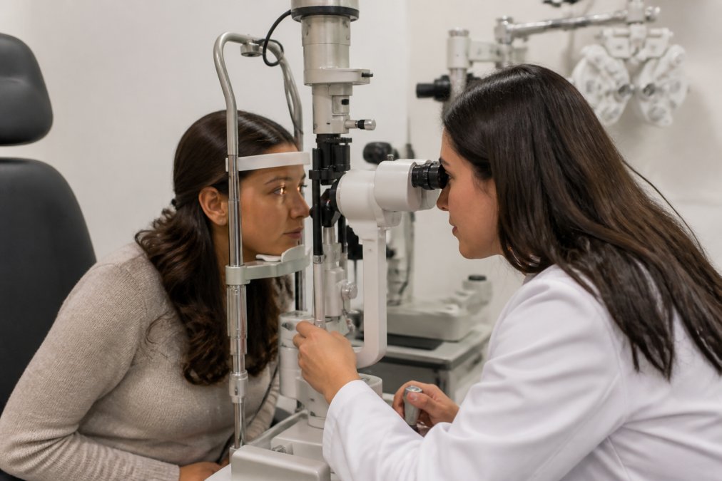
Dilated eye examination allows the ophthalmologist to visualize the back of your eye directly. Using a bright light and magnifying lens, they can inspect your retina, optic nerve, and blood vessels for signs of detachment, hemorrhage, artery blockage, or inflammation.
Optical coherence tomography (OCT) is a non-invasive imaging test creating detailed cross-sectional images of your retina. It’s particularly useful for detecting retinal detachment, swelling, or structural changes and takes minutes.

Fundus photography captures detailed photographs of your retina and optic nerve, providing a permanent record for comparison.
Intraocular pressure measurement (tonometry) checks the fluid pressure inside your eye. Elevated pressure can indicate acute glaucoma. This test is quick and painless.
Blood tests may be ordered if giant cell arteritis is suspected, particularly if you’re over 50 with scalp tenderness or jaw pain. Blood tests measure inflammatory markers like erythrocyte sedimentation rate (ESR) and C-reactive protein (CRP).
Carotid ultrasound or imaging may be recommended if artery occlusion is suspected, to check for blockages in the carotid arteries supplying blood to your eyes and brain.
Visual field testing may be performed to map exactly which areas of your vision are affected.
The entire emergency evaluation typically takes 1-2 hours. The goal is to reach a diagnosis quickly so treatment can begin without delay. For some conditions like retinal artery occlusion, treatment options exist only within a narrow time window (sometimes as little as 4-6 hours from symptom onset).
Recovery and Long-Term Outlook After Sudden Vision Loss
The outlook after sudden vision loss depends entirely on the cause, how quickly treatment began, and how much damage occurred before intervention.
Retinal artery occlusion outcomes vary widely. If treated within the critical window (ideally within 4-6 hours), some vision recovery is possible. However, if the blockage persists beyond this window, permanent vision loss in the affected area is likely.
Retinal detachment has a much better prognosis if caught early. Surgery to reattach the retina, performed urgently (ideally within days), can restore vision in many cases. Patients treated promptly often recover good vision; those with delayed treatment may have lasting blind spots.
Vitreous hemorrhage often resolves gradually as blood reabsorbs. Vision typically improves over weeks to months. Extensive or recurrent bleeding may require surgery.
Acute angle-closure glaucoma, when treated promptly with medications or laser procedures, often preserves vision. Delayed treatment can cause permanent optic nerve damage.
Giant cell arteritis requires prompt corticosteroid treatment to prevent vision loss in the other eye. Early treatment halts progression, though vision already lost is often not recovered.
Endophthalmitis requires aggressive antibiotic or antifungal therapy. Outcomes depend on how quickly treatment begins and the causative organism.
Patients who’ve experienced sudden vision loss often benefit from rehabilitation and psychological support. Low-vision rehabilitation specialists can teach adaptive techniques to maximize remaining vision. For some conditions like retinal detachment, the risk of recurrence in the other eye is elevated, warranting regular monitoring. For vascular events, managing underlying conditions like blood pressure, cholesterol, and diabetes reduces future risk.
Clear Vision San Antonio recommends that anyone who’s experienced sudden vision loss maintain close follow-up care with an ophthalmologist, even after initial treatment and recovery.
Sudden vision loss is a medical emergency demanding immediate action. Conditions causing it, retinal detachment, eye stroke, acute glaucoma, and others, can lead to permanent blindness if treatment is delayed. If you or someone in your care experiences sudden vision loss, go to the emergency room immediately. Early diagnosis and treatment offer the best chance of preserving vision. Clear Vision San Antonio is committed to helping patients understand eye health emergencies and providing expert care when vision is at stake. If you have concerns about your eye health or have experienced vision changes, request an appointment with our team to discuss your individual situation and ensure your vision is protected.
Frequently Asked Questions
What are the most common medical emergencies that cause sudden vision loss?
Retinal detachment, retinal artery occlusion (eye stroke), vitreous hemorrhage, acute glaucoma, and giant cell arteritis are among the most serious causes. Retinal detachment occurs when the light-sensitive tissue separates from the back of the eye and requires emergency surgery to prevent permanent damage. A retinal artery occlusion cuts off blood flow to the retina, causing sudden vision loss similar to a stroke. Each condition demands urgent evaluation by an ophthalmologist to preserve vision.
Is sudden vision loss in one eye considered a stroke symptom?
Sudden vision loss in one eye can indicate a retinal artery occlusion, also called an eye stroke, which is a medical emergency. This occurs when a blood clot or plaque blocks blood flow to the retina. Unlike a brain stroke affecting both eyes, an eye stroke affects only one eye and creates a sudden, painless loss of vision. Seek emergency medical attention immediately if you experience this symptom, as prompt treatment within hours may preserve remaining vision.
What should I do if I experience a sudden dark curtain over my vision?
A dark curtain or shadow moving across your field of vision is a classic sign of retinal detachment and requires emergency care. Do not wait or assume it will resolve. Call 911 or go to the nearest emergency room immediately. Retinal detachment is painless but progresses rapidly; delaying treatment can result in permanent vision loss. Lie flat if possible while waiting for help, and avoid rubbing or pressing on your eye.
How quickly should I be seen by an ophthalmologist for sudden vision loss?
Seek emergency care immediately, do not wait hours or until the next business day. Many causes of sudden vision loss, including retinal detachment, eye stroke, and acute glaucoma, can cause permanent damage within hours if untreated. The first few hours are critical for preserving vision. Go to an emergency room or call your ophthalmologist's emergency line right away. Prompt diagnostic evaluation and treatment can mean the difference between regaining vision and permanent blindness.
This article was written using GrandRanker