

Table of Contents
- Understanding Early Stage Glaucoma: The Silent Thief of Vision
- Recognizing Symptoms of Early Stage Glaucoma
- What Causes Early Stage Glaucoma: Risk Factors and Causes
- How Is Glaucoma Diagnosed: Testing and Screening
- Glaucoma Treatment Options: Managing Your Eye Health
- When to See an Ophthalmologist: Taking Action Early
- Lifestyle and Eye Health: Supporting Your Vision Long-Term
- Conclusion: Protect Your Vision Through Early Detection
Last Updated: July 6, 2026
Understanding Early Stage Glaucoma: The Silent Thief of Vision
Glaucoma affects millions worldwide, yet many don’t realize they have it until significant vision loss occurs. Early stage glaucoma progresses silently, with symptoms so subtle that people often miss them entirely. This guide covers what you need to know about recognizing early warning signs before irreversible damage happens.
The condition develops when intraocular pressure damages the optic nerve, the bundle of nerve fibers responsible for transmitting visual information to the brain. Unlike some eye conditions that announce themselves with pain or obvious vision changes, early stage glaucoma often progresses without symptoms. By the time patients notice changes, peripheral vision loss may already be advanced. Understanding the symptoms of early stage glaucoma is the difference between preserving your vision and losing it. Early detection and treatment can slow or halt progression, but only if you know what to watch for.
Why Early Detection Matters
Detecting glaucoma before significant optic nerve damage occurs is the single most important factor in preserving vision long-term. Once the optic nerve sustains damage, that loss is permanent. The optic nerve cannot regenerate, which means treatment focuses entirely on preventing further deterioration rather than restoring what’s been lost.
Research shows that people diagnosed and treated in early stages maintain better visual function throughout their lives compared to those diagnosed after substantial damage has occurred. Early detection also opens treatment options that work best when started sooner. Medications are more effective at controlling intraocular pressure in early disease, and surgical interventions tend to produce better outcomes when performed before advanced damage develops.
Schedule a comprehensive eye exam every 1-2 years if you have no risk factors, and annually if you’re over 40 or have family history of glaucoma.
Recognizing Symptoms of Early Stage Glaucoma
Subtle Vision Changes
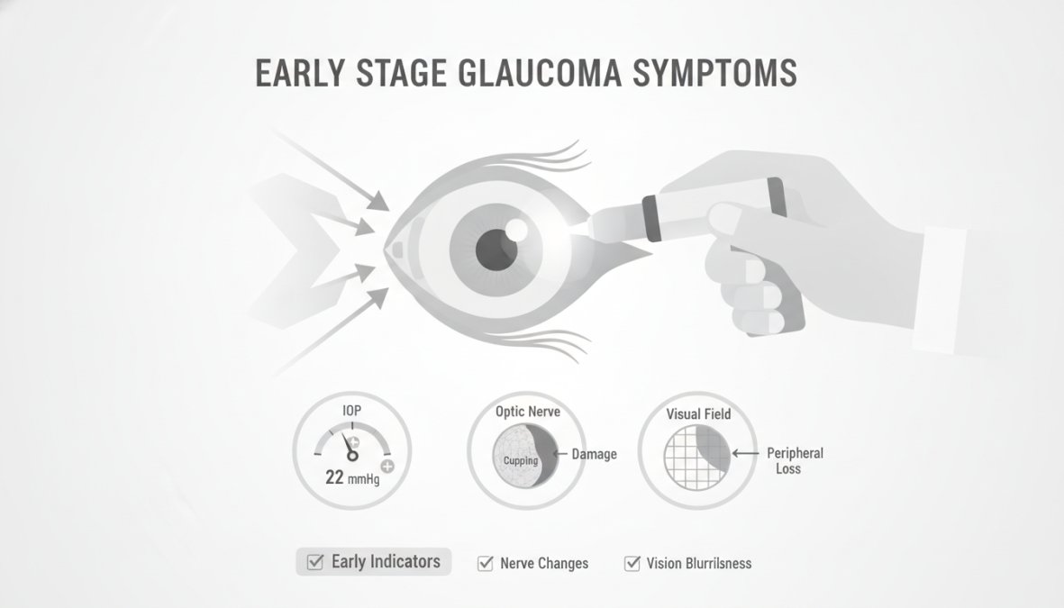
The early signs of vision changes are often so gradual that people attribute them to normal aging or assume they need glasses adjusted. Many describe noticing that their peripheral vision, the ability to see objects to the side without moving their eyes, seems slightly narrower. Some patients report difficulty with night driving or vision that seems slightly hazier overall, though the haziness doesn’t match what new glasses prescriptions can correct.
Blurred vision can occur, though it’s usually mild and intermittent rather than constant. These changes develop over months or years, making them easy to overlook.
Physical and Visual Discomfort
Eye pain is not typically associated with early stage glaucoma. Most people with early glaucoma experience no pain whatsoever. The absence of pain is actually part of why glaucoma is so dangerous; patients don’t feel anything wrong.
However, some people report mild eye discomfort or a sensation of pressure behind the eye, particularly in the morning. This pressure sensation differs from pain, it’s described as a dull feeling rather than sharp pain. Halos around lights, particularly visible at night around streetlights or headlights, can occasionally occur in early stages. These halos appear as rainbow-colored rings around bright light sources.
If you experience sudden eye pain, sudden vision loss, severe headache with vision changes, or nausea with eye discomfort, seek immediate medical attention. These can indicate acute angle-closure glaucoma, which requires emergency treatment.
What Causes Early Stage Glaucoma: Risk Factors and Causes
Elevated Intraocular Pressure
Intraocular pressure, the fluid pressure inside the eye, is the primary modifiable risk factor for glaucoma. The eye produces fluid called aqueous humor that maintains the eye’s shape and nourishes internal structures. When the eye’s drainage system doesn’t function properly, fluid accumulates and pressure builds.
Normal intraocular pressure ranges from 12 to 22 millimeters of mercury (mmHg). However, some people develop glaucoma at pressures within this "normal" range, while others tolerate higher pressures without damage. Elevated intraocular pressure damages the optic nerve by restricting blood flow to the nerve fibers. This damage is progressive and cumulative.
Genetics play a significant role in intraocular pressure regulation. Age also matters; intraocular pressure tends to increase slightly with age. Certain medications, particularly corticosteroids, can raise intraocular pressure in susceptible individuals.
Genetic and Demographic Risk Factors
Family history is one of the strongest predictors of glaucoma risk. If your parent, sibling, or grandparent has glaucoma, your risk increases substantially. Age significantly increases glaucoma risk. While glaucoma can develop at any age, it becomes increasingly common after age 40.
Race and ethnicity influence glaucoma risk. African Americans develop glaucoma more frequently and at earlier ages than other groups, often with more aggressive disease progression. Hispanic populations also show higher prevalence than non-Hispanic white populations. Asian populations have higher rates of angle-closure glaucoma specifically.
Medical conditions contribute to glaucoma risk. Diabetes increases risk, as does high blood pressure. Severe myopia (nearsightedness) is associated with higher glaucoma risk. Thyroid eye disease and previous eye injuries can elevate intraocular pressure.
If you have multiple risk factors, family history, age over 40, high eye pressure, or certain medical conditions, prioritize regular [eye exams](/managing-diabetic-retinopathy-through-eye-exams/).
How Is Glaucoma Diagnosed: Testing and Screening
Comprehensive Eye Screening Process
A comprehensive eye exam is the foundation of glaucoma detection. Your eye care professional begins by reviewing your medical history, including family history of glaucoma, previous eye injuries, medications, and systemic diseases. Visual acuity testing measures how clearly you see at various distances.
During the dilated exam, your eye care professional carefully examines the optic disc, the point where the optic nerve enters the eye. They assess the appearance of the nerve tissue and look for characteristic changes associated with glaucoma. The optic cup, a natural depression in the center of the optic disc, may enlarge as glaucoma progresses.
Intraocular pressure measurement, called tonometry, is essential. Applanation tonometry uses a small probe to gently flatten the cornea and measure pressure. Non-contact tonometry uses a puff of air and requires no contact with the eye. Both methods are accurate when performed properly.
Advanced Diagnostic Tools
Visual field testing, also called perimetry, maps your complete field of vision by testing your ability to see points of light at various locations and intensities. Early glaucoma typically causes peripheral vision loss in specific patterns. Automated perimetry is the standard method, where you sit before a dome-shaped instrument and respond to light flashes.
Optical coherence tomography (OCT) provides detailed cross-sectional images of the optic nerve and retinal nerve fiber layer. This technology measures the thickness of nerve fiber layers, which thin as glaucoma progresses. OCT detects subtle structural changes before functional vision loss becomes apparent on visual field testing.
Gonioscopy examines the angle where the iris meets the cornea, the space through which fluid drains from the eye. This examination is crucial for distinguishing open-angle glaucoma from angle-closure glaucoma, as treatment differs significantly.
Pachymetry measures corneal thickness. Thinner corneas may give falsely low pressure readings, while thicker corneas may give falsely high readings. Knowing your actual corneal thickness allows your eye care provider to interpret pressure measurements more accurately.
| Diagnostic Test | What It Measures | When It’s Most Useful |
|---|---|---|
| Tonometry | Intraocular pressure | Initial screening and monitoring |
| Visual field testing | Peripheral vision function | Detecting early functional loss |
| OCT imaging | Optic nerve and nerve fiber layer thickness | Detecting structural changes early |
| Gonioscopy | Drainage angle configuration | Determining glaucoma type |
| Dilated exam | Optic disc appearance | Assessing nerve health directly |
Glaucoma Treatment Options: Managing Your Eye Health
Medical and Surgical Interventions
Medication is typically the first treatment approach for early stage glaucoma. Prostaglandin analogs, eye drops that increase fluid outflow from the eye, are often prescribed as initial therapy. These medications are applied once daily, usually in the evening, and are effective at lowering intraocular pressure.
Beta blockers and alpha-adrenergic agonists represent alternative medication classes that reduce intraocular pressure through different mechanisms. Carbonic anhydrase inhibitors also decrease fluid production. Your ophthalmologist selects medications based on your specific pressure levels, overall health, and potential side effects.
Medication adherence is critical but challenging. Many people with early glaucoma feel fine and struggle to remember daily eye drops. Setting phone reminders or linking drop administration to daily routines helps improve adherence.
Selective laser trabeculoplasty (SLT) targets the eye’s drainage system, stimulating it to function more efficiently. This in-office procedure takes about 10 minutes and can reduce intraocular pressure by 20-30% in many patients. SLT can be repeated if pressure rises again over time.
Conventional surgery, called trabeculectomy, creates a new drainage pathway for fluid to exit the eye. This procedure is typically reserved for cases where medications and laser treatment haven’t achieved adequate pressure control. Minimally invasive glaucoma surgeries (MIGS) represent newer approaches that create micro-drainage pathways with less risk than traditional surgery while still effectively lowering pressure.
When to See an Ophthalmologist: Taking Action Early
Schedule an appointment with an ophthalmologist, not just an optometrist, if you have any risk factors for glaucoma. Ophthalmologists have additional training in diagnosing and treating glaucoma specifically and can perform gonioscopy and interpret complex imaging results.
Seek immediate attention if you experience sudden vision changes, severe eye pain, headache with vision changes, or nausea accompanied by eye discomfort. These symptoms can indicate acute angle-closure glaucoma, a medical emergency requiring same-day treatment.
If you’ve already been diagnosed with glaucoma, regular follow-up appointments are essential. Early stage glaucoma typically requires eye exams every 3-6 months initially to ensure medications are controlling pressure adequately. Once stable, appointments might space to every 6-12 months. Visual field testing and imaging are usually repeated annually to monitor for progression.
Don’t assume that normal eye pressure means you don’t have glaucoma. Some people develop glaucoma despite normal-range pressures, a condition called normal-tension glaucoma. If you have risk factors or family history, comprehensive evaluation is warranted even if pressure readings seem acceptable.
If cost is a barrier to regular eye care, many ophthalmology practices offer payment plans or can refer you to community health centers providing discounted services.
Lifestyle and Eye Health: Supporting Your Vision Long-Term
While medications and surgical interventions are the primary treatments for glaucoma, lifestyle factors support overall eye health. Regular aerobic exercise, such as brisk walking, swimming, or cycling, may help reduce intraocular pressure modestly and improves blood flow to the optic nerve.
Diet rich in antioxidants supports eye health. Foods containing lutein, zeaxanthin, and vitamins C and E may protect against age-related eye disease. Leafy greens, colorful vegetables, nuts, and fish are particularly beneficial. Omega-3 fatty acids from fish or supplements may support optic nerve health.
Avoid activities that significantly elevate intraocular pressure. Inverted yoga poses, heavy weightlifting, and straining during bowel movements can temporarily raise eye pressure. Manage systemic conditions that affect glaucoma risk. Blood pressure control reduces glaucoma progression risk. Diabetes management prevents complications that worsen glaucoma.
Sleep position may matter for some people. Sleeping with the head elevated slightly, using an extra pillow, may help maintain more stable intraocular pressure throughout the night. Stress management supports overall health and may influence intraocular pressure. Meditation, deep breathing exercises, and activities you enjoy reduce stress and promote general wellbeing.
Limit caffeine intake modestly. While caffeine doesn’t cause glaucoma, some studies suggest high caffeine consumption may slightly elevate intraocular pressure in susceptible individuals. Moderate consumption, one to two cups of coffee daily, is generally considered safe.
Conclusion: Protect Your Vision Through Early Detection
Early stage glaucoma remains the leading cause of irreversible vision loss in adults, yet most cases are preventable through early detection and treatment. The challenge isn’t that glaucoma is untreatable, it’s that many people don’t know they have it until significant damage has occurred. Regular eye exams, particularly for those with risk factors, are your strongest defense against vision loss.
Clear Vision San Antonio specializes in comprehensive glaucoma evaluation and management, combining advanced diagnostic technology with personalized treatment planning. Our ophthalmologists use OCT imaging, visual field testing, and gonioscopy to detect early changes before vision is affected.
Frequently Asked Questions
What are the first signs of glaucoma?
Early symptoms of early stage glaucoma are often absent, which is why it's called the 'silent thief of sight.' When symptoms do appear, they may include subtle peripheral vision loss, mild eye discomfort, or halos around lights. Some individuals experience blurred vision or eye redness. Regular eye screening with tonometry and visual field testing can detect elevated intraocular pressure and optic nerve damage before noticeable vision loss occurs.
Does early stage glaucoma have any symptoms?
Most people with early stage glaucoma experience no symptoms at all. This is the critical challenge, the disease progresses silently, gradually damaging the optic nerve and retinal nerve fiber layer without causing pain or obvious vision changes. This is why routine screening is essential, particularly for those with family history, elevated eye pressure, or other risk factors. Early detection through dilated eye exams and visual field tests can identify the condition before significant vision loss occurs.
How is early stage glaucoma diagnosed?
Early stage glaucoma is diagnosed through comprehensive eye screening by an ophthalmologist or optometrist. Key diagnostic tools include tonometry (measuring intraocular pressure), visual field testing (assessing peripheral vision), optical coherence tomography (OCT) imaging of the optic disc and retinal nerve fiber layer, and dilated eye exams. These tests detect subtle changes in eye pressure and optic nerve damage before symptoms develop, making early detection possible and preventing progression to tunnel vision or blindness.
Can you reverse early stage glaucoma?
Early stage glaucoma cannot be reversed, but it can be managed effectively when caught early. Glaucoma treatment options focus on slowing or halting disease progression by lowering intraocular pressure through medication, laser procedures, or surgery. Starting treatment early, before significant optic nerve damage occurs, is crucial for preserving vision long-term. Regular monitoring and adherence to prescribed treatment help prevent the condition from advancing to later stages with permanent vision loss.
Vision loss from glaucoma is permanent, but progression is preventable with early intervention. Don’t wait for symptoms to develop, symptoms of early stage glaucoma are often imperceptible until irreversible damage has occurred. Request an appointment with Clear Vision San Antonio today for a comprehensive glaucoma evaluation that protects your vision for life.
This article was written using GrandRanker