
Table of Contents
- Why Vision Changes After Forty: Understanding the Natural Aging Process
- Presbyopia Symptoms and Near Vision Challenges
- Common Vision Changes After 40 You Should Know
- Dry Eye Syndrome Treatment and Management
- Age-Related Eye Conditions: Cataracts, Glaucoma, and Macular Degeneration
- Night Driving Vision Problems: Causes and Solutions
- Comprehensive Eye Exam Importance for Early Detection
- Nutritional Impact on Eye Health and Vision Aging
Last Updated: June 20, 2026
Understanding why vision changes after forty is one of the most common concerns patients bring to eye care appointments. The eyes undergo real, measurable structural shifts starting in the fourth decade of life, and most people are caught off guard when they suddenly struggle to read a restaurant menu. Below, we break down the biology, the warning signs, and the treatment options.
Why Vision Changes After Forty: Understanding the Natural Aging Process
Presbyopia is the primary reason why vision changes after forty, and it is not a disease. Presbyopia is the gradual loss of the eye’s ability to focus on near objects, caused by the natural stiffening of the crystalline lens inside the eye. This process is universal, nobody escapes it, regardless of prior vision quality.
How the Lens Loses Flexibility with Age
The crystalline lens sits just behind the pupil and works like a camera autofocus system. Tiny ciliary muscles surrounding it contract and relax to change its shape, allowing the eye to shift focus between near and distant objects. This process is called accommodation.
Before age 40, the lens is soft and pliable. After 40, the lens proteins gradually harden, reducing the lens’s ability to change curvature. The ciliary muscles are still working fine, the problem is that the lens no longer responds to their signals with the same flexibility. According to the American Academy of Ophthalmology’s patient education resources, the amplitude of accommodation drops steadily from childhood onward, with the most noticeable functional impact typically occurring between ages 40 and 45. This process has been happening since your early twenties; by 40, the reserve capacity is gone.
Presbyopia is not a sudden failure. It is the final stage of a decades-long process. Understanding this helps patients set realistic expectations for treatment timelines and outcomes.
Presbyopia Symptoms and Near Vision Challenges
The classic symptom pattern is specific: you hold your phone or book at arm’s length to bring text into focus. Headaches after reading are common, and dim restaurants become genuinely difficult.
Reading Glasses, Bifocals, and Progressive Lenses
Reading glasses add magnifying power for near distances and work well for people with no prior refractive error, though they require constant removal for distance tasks. Bifocals divide the lens into two zones but create an abrupt visual jump that some patients never fully adapt to.
Progressive lenses are the current standard, providing a gradient of correction from distance at the top through intermediate in the middle to near at the bottom, with no visible line. Adaptation typically takes two to four weeks. The right choice depends on lifestyle: simple reading glasses suit those who primarily need near vision help, while progressives benefit those needing both distance and near correction.
Common Vision Changes After 40 You Should Know
Several changes happen in parallel with presbyopia, and conflating them leads to frustration when reading glasses alone do not solve every problem.
Blurred Vision, Faded Colors, and Reduced Night Vision
The lens yellows with age, filtering out blue light and causing colors to appear muted. Night vision deteriorates because the pupil becomes less responsive and opens less fully in low light, the retina becomes less sensitive, and contrast sensitivity drops. These are normal aging changes, but they can also signal cataracts or other conditions requiring professional evaluation.
Digital Eye Strain and Light Sensitivity
Digital eye strain has become one of the most common complaints among adults over 40. Screens demand sustained near focus, and aging eyes have less accommodative reserve. Light sensitivity increases with age because the aging lens scatters light more than a clear, young lens does, also contributing to halos and glare around lights at night.
Do not dismiss increasing light sensitivity or halos as simple aging. These symptoms can indicate early cataract formation or elevated intraocular pressure. An ophthalmologist should evaluate them before attributing them to normal aging alone.
Dry Eye Syndrome Treatment and Management
Dry eye syndrome is one of the most underdiagnosed conditions in patients over 40. The lacrimal glands produce fewer tears with age, and the lipid layer of the tear film thins, causing tears to evaporate faster.
Treatment typically follows a stepwise approach. Artificial tears are the starting point, with preservative-free formulations preferred for frequent use. Warm compresses and lid hygiene address meibomian gland dysfunction, responsible for the majority of dry eye cases in this age group. Omega-3 supplementation has supporting evidence for improving tear film quality. For moderate to severe cases, prescription anti-inflammatory drops or punctal plugs are effective next steps. According to the National Eye Institute’s dry eye overview, dry eye affects a substantial portion of adults over 50, with women experiencing higher rates due to hormonal changes.
Age-Related Eye Conditions: Cataracts, Glaucoma, and Macular Degeneration
Normal aging changes are manageable. Age-related eye diseases require medical intervention. The three most significant are cataracts, glaucoma, and macular degeneration.
Cataracts and Lens Clouding Explained
A cataract is a clouding of the crystalline lens, caused by the same protein aggregation that drives presbyopia, but progressing further. The lens becomes opaque rather than merely stiff, producing blurred vision that worsens in bright light, increased glare and halos at night, faded colors, and progressive difficulty with visual acuity.
Cataract surgery is the most commonly performed surgical procedure in the United States and is highly effective. The clouded lens is removed and replaced with an intraocular lens implant. Advanced lens implants can correct presbyopia simultaneously, reducing or eliminating the need for reading glasses after surgery.
Glaucoma Risks and Intraocular Pressure
Glaucoma is a group of conditions that damage the optic nerve, most commonly through elevated intraocular pressure. Damage to the optic nerve is irreversible, making glaucoma particularly dangerous: it typically causes no symptoms until significant vision loss has already occurred.
Risk increases substantially after age 40 and further after 60. Family history, elevated intraocular pressure, thin corneas, and certain ethnic backgrounds all increase risk. Intraocular pressure screening is a standard component of comprehensive eye exams and is the primary detection tool. Treatment focuses on reducing intraocular pressure through eye drops, laser therapy, or surgery, depending on severity.
Macular Degeneration and Retinal Changes
Age-related macular degeneration (AMD) affects the macula, the central portion of the retina responsible for sharp, detailed vision. It is the leading cause of vision loss in adults over 50 in developed countries.
AMD presents in two forms. Dry AMD involves gradual thinning of the macula and accumulation of drusen deposits. Wet AMD involves abnormal blood vessel growth beneath the retina, which can cause rapid and severe central vision loss. Early dry AMD may produce no noticeable symptoms, reinforcing the importance of regular retinal examination. Risk factors include smoking, cardiovascular disease, family history, and prolonged UV exposure.
Night Driving Vision Problems: Causes and Solutions
Night driving vision problems result from reduced pupil dilation, decreased contrast sensitivity, and increased lens scatter. Oncoming headlights cause more glare, road markings are harder to distinguish, and reaction time to obstacles slows.
Practical solutions include anti-reflective coatings on lenses, which significantly reduce glare from oncoming lights. Regular glasses prescriptions should be updated, as even small uncorrected refractive errors are amplified in low-contrast night conditions. Patients with cataracts often report dramatic improvement in night driving after surgery.
Request an anti-reflective coating specifically optimized for nighttime driving when ordering new lenses. Premium versions designed for night conditions provide noticeably better performance for patients who drive frequently after dark.
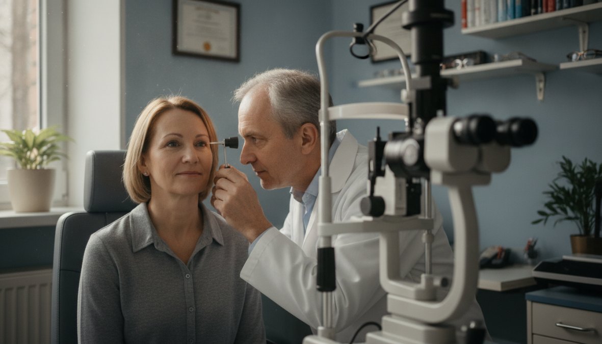
Comprehensive Eye Exam Importance for Early Detection
The single most effective intervention available for protecting vision after 40 is a comprehensive eye exam on a consistent schedule. This is not a vision screening, a vision screening tests only visual acuity. A comprehensive exam evaluates the entire eye, including structures that show disease long before symptoms appear.
What to Expect During an Eye Exam with an Ophthalmologist
A comprehensive exam includes dilation to allow direct visualization of the retina, optic nerve, and blood vessels. The ophthalmologist examines the lens for cataract formation, the cornea for changes in clarity and shape, and the vitreous for signs of detachment or floaters. Patient history matters significantly, as medications, family history, systemic conditions like diabetes and hypertension, and recent visual symptoms all inform the examination.
Visual Acuity Testing and Intraocular Pressure Screening
Visual acuity testing establishes your baseline corrected and uncorrected vision. Intraocular pressure measurement screens for glaucoma risk. Optical coherence tomography, when indicated, produces high-resolution cross-sectional images of the retina and optic nerve that can detect early AMD and glaucoma changes.
Adults over 40 with no known risk factors should have a comprehensive exam every one to two years. Those with risk factors should follow a schedule recommended by their ophthalmologist, often annually or more frequently.
Nutritional Impact on Eye Health and Vision Aging
Diet is one of the few modifiable factors that genuinely influences the rate of age-related eye disease progression. Lutein and zeaxanthin are carotenoids concentrated in the macula that filter high-energy blue light and act as antioxidants, found in leafy greens, eggs, and corn. Omega-3 fatty acids support tear film quality and reduce dry eye severity. Vitamins C and E, zinc, and copper have demonstrated benefit in slowing AMD progression in patients with intermediate disease. Smoking is the single most damaging lifestyle factor for AMD risk and should be avoided entirely.
Self-Assessment Checklist: Monitoring Your Vision After 40
Use this checklist to track changes between professional exams. Any item marked yes warrants a conversation with an ophthalmologist.
- Difficulty reading small print without holding it farther away
- Eye strain or headaches after reading or screen use
- Blurred vision that fluctuates throughout the day
- Increased sensitivity to glare or bright light
- Halos or starbursts around lights at night
- Colors appearing less vivid or slightly yellowed
- Difficulty driving after dark
- Straight lines appearing wavy or distorted (potential AMD symptom, seek evaluation promptly)
- Sudden floaters or flashes of light (seek urgent evaluation)
- Dry, gritty, or burning eye sensation daily
This checklist is not a diagnostic tool. It is a structured way to notice and communicate changes to your eye care provider before they become significant problems.
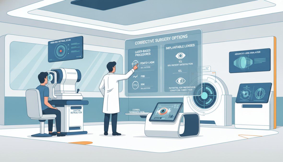
Technological Advancements in Corrective Surgery and Vision Correction
The options available for correcting age-related vision changes in 2026 extend well beyond reading glasses. Corrective surgery has advanced substantially.
(/best-vision-correction-for-over-50/)]
LASIK remains the most widely performed refractive procedure for correcting nearsightedness, farsightedness, and astigmatism. Monovision LASIK corrects one eye for distance and one for near. ICL, or implantable collamer lens, is an alternative for patients whose corneal thickness or prescription falls outside LASIK’s range.
For presbyopia specifically, premium intraocular lens technology offers significant advances. Extended depth-of-focus lenses and trifocal lenses provide functional vision at near, intermediate, and distance ranges simultaneously, often reducing dependence on glasses to a minimal level. These are most commonly placed during cataract surgery but can also be implanted through refractive lens exchange in patients who have not yet developed cataracts.
Vision after 40 changes in ways that are predictable, treatable, and in many cases preventable from progressing to serious disease. Request a comprehensive eye exam and get a complete picture of your eye health before problems develop.
Frequently Asked Questions
Why does vision deteriorate after 40?
Vision changes after 40 due to the natural aging process. The lens in your eye loses flexibility over time, making it harder to focus on near objects, a condition called presbyopia. Additionally, the pupil becomes smaller, reducing light entry, and the retina becomes less sensitive. Other age-related changes include increased lens clouding (cataracts), reduced tear production causing dry eyes, and changes in the cornea. These combined factors result in presbyopia symptoms like blurred near vision, reduced night vision, and difficulty with digital eye strain.
What is the most common vision change after 40?
Presbyopia is the most common vision change after 40. It affects nearly everyone and occurs because the lens loses its flexibility, making it difficult to focus on near objects like text. This is why many people begin needing reading glasses or bifocals around age 40-45. Unlike other age-related conditions, presbyopia is a natural part of the aging process and not a disease. It can be corrected with reading glasses, progressive lenses, bifocals, or corrective surgery options like LASIK.
When should I see an eye doctor for vision changes after 40?
You should schedule a comprehensive eye exam immediately if you notice sudden vision changes, significant blurred vision, eye pain, or light sensitivity. Even without symptoms, the American Academy of Ophthalmology recommends annual eye exams after 40 to screen for conditions like glaucoma, cataracts, and macular degeneration. Early detection through visual acuity testing and intraocular pressure screening can prevent serious vision loss. An ophthalmologist can assess your focusing ability and recommend appropriate corrective solutions.
Can I improve my vision after 40 without glasses?
While presbyopia cannot be reversed, several options exist beyond traditional reading glasses. Technological advancements in corrective surgery, such as LASIK and advanced lens implants, can help restore clear vision at near, far, and intermediate distances. Nutritional support with antioxidants may slow age-related decline. However, most people over 40 will need some form of vision correction. A comprehensive eye exam with an ophthalmologist can determine which solutions, from progressive lenses to surgical options, best suit your lifestyle and visual needs.
This article was written using GrandRanker