

Quick Summary
Diabetic retinopathy treatment depends mainly on the disease stage and whether macular swelling is present. Options include anti-VEGF or steroid injections, focal or panretinal laser, and vitrectomy surgery for advanced complications. Early-stage disease is often monitored closely while blood sugar and blood pressure are controlled.
- Stage, not age or years with diabetes, usually determines which treatment is considered first.
- A dilated comprehensive eye exam is the standard way to detect diabetic retinopathy.
- Anti-VEGF injections are often used for diabetic macular edema and proliferative disease.
- Panretinal laser may reduce side or night vision, a trade-off doctors weigh carefully.
- Vitrectomy is considered for non-clearing vitreous hemorrhage or tractional retinal detachment.
Table of Contents
- How Diabetic Retinopathy Progresses and Why Stage Decides Treatment
- Diabetic Eye Exam: How Retinopathy Is Detected and Monitored
- Diabetic Retinopathy Injections: Anti-VEGF and Steroid Options
- Laser Treatment for Diabetic Retinopathy: When Photocoagulation Is Used
- Vitrectomy for Diabetic Retinopathy: Surgery for Advanced Disease
- Side-by-Side Comparison, Risks, Recovery and Cost in San Antonio
- Booking Your Retina Evaluation in San Antonio
- Frequently Asked Questions
How Diabetic Retinopathy Progresses and Why Stage Decides Treatment
Diabetic retinopathy is damage to the blood vessels of the retina caused by long-term high blood sugar (Diabetic Retinopathy). The stage of the disease, not the patient’s age or the number of years they have had diabetes, is what usually determines which diabetic retinopathy treatment is considered first.
Most people with diabetic retinopathy have no symptoms in the early stages (Diabetic Retinopathy). Vision can look normal while small blood vessels leak or close off, which is why routine screening matters so much for people with diabetes.
Nonproliferative vs. Proliferative Disease
Diabetic retinopathy is generally described in two broad categories. In nonproliferative disease, weakened vessels bulge, leak fluid, or become blocked. This stage is often monitored closely rather than treated right away, with the goal of tightening blood sugar and blood pressure control and checking the retina regularly.
Proliferative disease is more advanced. The retina responds to poor blood flow by growing new, fragile blood vessels, which can bleed into the eye or pull on the retina. This stage carries a higher risk of significant vision loss and usually calls for prompt treatment.
Diabetic Macular Edema: The Swelling That Blurs Central Vision
Diabetic macular edema (DME) occurs when fluid collects in the macula, the central part of the retina responsible for sharp detail. For a broader look at treatment options for macula-related disease, including wet and dry forms, see our complete guide. It can develop at any stage of retinopathy and is one of the most common causes of blurred central vision in people with diabetes.
Because DME affects reading and recognizing faces, patients often notice it first. Treatment choices for DME, including injections and laser, are covered in the sections below.
Diabetic Eye Exam: How Retinopathy Is Detected and Monitored
A dilated comprehensive eye exam is the standard way to detect diabetic retinopathy. Drops widen the pupil so the eye doctor can examine the retina, and imaging such as optical coherence tomography (OCT) can measure swelling in the macula. Many people with diabetes are advised to have a dilated exam at least once a year, though the schedule can be shorter for people with existing disease. Your eye doctor will recommend the right interval for you.
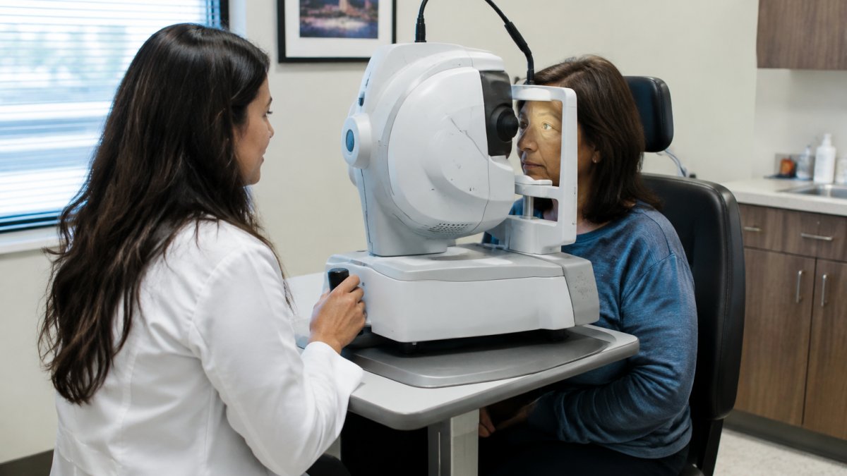
Detection is only the first step. Results guide decisions about monitoring, medication changes, and whether treatment is needed. For a closer look at screening frequency and what to expect, see our guide to managing diabetic retinopathy through eye exams. You can also read our diabetic eye exam frequency guide for a breakdown of how often different patients should be screened.
Regular imaging is especially useful because early changes are not always visible to the patient. American Academy of Ophthalmology diabetic retinopathy guidance recommends a dilated exam as the baseline test for people with diabetes. To understand why imaging is central to detection, read our article on why OCT scans are essential for early eye disease detection.
Diabetic Retinopathy Injections: Anti-VEGF and Steroid Options
Injections place medication directly into the eye and are among the most widely used diabetic retinopathy treatment options for diabetic macular edema and proliferative disease. The two main categories are anti-VEGF drugs, which block a protein that drives abnormal blood vessel growth and leakage, and corticosteroids, which reduce inflammation and swelling.
Anti-VEGF injections are one option used for DME and proliferative disease. Patients usually need repeated injections on a schedule set by their doctor. Steroid implants or injections may be used when anti-VEGF therapy is not suitable or does not fully control swelling. Side effects can include eye pressure changes and cataract development with steroids.
The experience of an injection is brief. The eye is numbed, and most patients describe pressure rather than sharp pain. Your doctor can explain how the schedule and risks apply to your specific case.
Laser Treatment for Diabetic Retinopathy: When Photocoagulation Is Used
Laser photocoagulation uses focused light to seal leaking vessels or to shrink areas of the retina that are not getting enough oxygen. Panretinal laser is used for proliferative disease, while focal or grid laser may be used for DME.
Laser can slow progression in some cases, but it does not restore vision that has already been lost. Some patients notice reduced side vision or night vision after panretinal treatment, which is a trade-off doctors weigh carefully. Laser is often combined with injections, depending on the stage and the patient’s response.
Patients usually go home the same day. Mild discomfort, light sensitivity, or temporary blurriness may occur for a short time. Ask your provider about the expected recovery for your treatment.
Vitrectomy for Diabetic Retinopathy: Surgery for Advanced Disease
A vitrectomy is a surgical procedure that removes the vitreous gel from inside the eye.
When surgery is considered. A common pattern is to wait a period of weeks to months for a vitreous hemorrhage to clear spontaneously, because many do.
What the procedure involves. The surgeon makes small incisions in the eye and uses fine instruments to remove the vitreous gel, clear blood, and peel scar tissue off the retina.
Recovery and follow-up. Recovery is longer than for injections or laser. Patients often use medicated eye drops for several weeks, may need to maintain a specific head position if a gas bubble is used, and are usually advised to avoid strenuous activity and air travel while the bubble is present.
Risks. As with any eye surgery, risks include infection, bleeding, retinal detachment, cataract, and increased eye pressure. Some patients need a second surgery. Surgery does not cure diabetes, so ongoing blood sugar and blood pressure control remain essential to protecting the retina afterward.
Urgent warning signs. Seek prompt evaluation for sudden vision loss, a sudden increase in floaters, flashes of light, or a curtain-like shadow across the vision. These can signal a hemorrhage or retinal detachment and are reasons not to wait for a routine appointment.
Questions worth asking your surgeon. What is the goal of surgery in my case, clearing blood, reattaching the retina, or both? Will a gas bubble or silicone oil be used, and what restrictions does that create? How long until I can drive, fly, or return to work? What is the plan if a second procedure is needed?
Side-by-Side Comparison, Risks, Recovery and Cost in San Antonio
The table below summarizes the main options by what each is trying to accomplish, how often it is typically delivered, and what recovery usually looks like. The right choice depends on your diagnosis, stage, whether macular edema is present, your prior treatment history, and your overall health, not on which option sounds strongest in isolation.
| Treatment | Primary goal | Typically used for | Typical schedule | Main advantages | Main drawbacks | Recovery and follow-up |
|---|---|---|---|---|---|---|
| Anti-VEGF injections | Reduce leakage and abnormal vessel growth | Diabetic macular edema; proliferative disease | Repeated injections on a doctor-set interval, often every 4-8 weeks initially, then extended based on response | Can improve or stabilize vision; often first-line for center-involving edema | Frequent visits; small risk of infection, inflammation, or pressure spike | Same-day return to most activities; avoid rubbing the eye; report pain, redness, or vision loss promptly |
| Steroid injections or implants | Reduce inflammation and swelling | Diabetic macular edema when anti-VEGF is insufficient or unsuitable | Single injection or sustained-release implant; repeat based on duration and response | Longer-acting options; useful when anti-VEGF alone does not control swelling | Raises eye pressure; cataract risk; small infection risk | Same-day return; pressure checks and cataract monitoring at follow-up |
| Focal or grid laser | Seal leaking vessels and reduce edema | Some cases of diabetic macular edema, especially non-center-involving | Usually one session, sometimes repeated | Durable reduction in leakage | Less effective for center-involving edema; may cause scarring | Same day; mild light sensitivity or temporary blurriness |
| Panretinal photocoagulation | Shrink oxygen-starved peripheral retina to reduce new vessel growth | Proliferative disease, especially high-risk features | Often divided into sessions | Slows progression and lowers severe vision loss risk | May reduce side or night vision; can worsen edema | Same day; mild discomfort and light sensitivity |
| Vitrectomy | Remove blood or scar tissue and reattach the retina | Non-clearing vitreous hemorrhage; tractional retinal detachment | One surgery, sometimes combined with other treatment | Addresses advanced complications that medications and laser cannot | Surgical risks including infection, bleeding, cataract, and pressure changes | Weeks of positioning restrictions and drops; vision may take months to stabilize |
How the decision is actually made. Stage is the starting point, but it is not the whole story. A patient with nonproliferative disease and no macular edema may be monitored with tighter blood sugar and blood pressure control rather than treated. A patient with center-involving diabetic macular edema is usually offered anti-VEGF therapy first, with steroids considered when response is incomplete or visits are difficult to sustain. Proliferative disease with high-risk features generally calls for prompt panretinal laser, often combined with anti-VEGF.
Watch out: Presenting these treatments as interchangeable is the most common mistake. A laser that is appropriate for peripheral new vessels is not a substitute for an injection that treats macular swelling, and a vitrectomy is not an upgrade from injections, it solves a different problem.
Cost, insurance, and access. Costs depend on the treatment, the number of visits, imaging, and your plan design. Ask the office for a good-faith estimate before scheduling. Coverage rules, prior authorization, and referral requirements differ by plan, so check with your insurer. Ask your insurer three questions before treatment: whether the specific drug or procedure is covered, whether prior authorization is required, and what your copay or coinsurance will be per visit.
Clear Vision San Antonio provides ophthalmological care for patients in San Antonio. Pricing depends on your individual needs, so please contact our office for current fees and insurance details.
Booking Your Retina Evaluation in San Antonio
If you have diabetes and have not had a recent dilated eye exam, or if you have noticed blurred, distorted, or dark spots in your vision, schedule an evaluation promptly. Early detection gives the widest range of treatment options.
Start by asking your primary care doctor or diabetes care team for a referral, then book an appointment with an eye doctor experienced in retinal disease. At your visit, bring a list of your medications, your most recent blood sugar results, and any questions about treatment, cost, or recovery.
At Clear Vision San Antonio, we provide personalized attention to each patient. Request an appointment to discuss your diabetic eye care with our team.
Frequently Asked Questions
What is the most effective treatment for diabetic retinopathy?
No single treatment fits every patient. The right option depends on the stage of disease, whether diabetic macular edema is present, and how the retina responds. Anti-VEGF injections are often used when swelling affects central vision, laser photocoagulation is used to slow abnormal blood vessel growth and reduce bleeding risk, and vitrectomy is reserved for advanced disease such as non-clearing vitreous hemorrhage or retinal detachment. Blood sugar and blood pressure control remain essential alongside any procedure. A retina specialist can explain which option fits your eyes.
How much do diabetic retinopathy injections typically cost?
Clear Vision San Antonio has not published pricing for this service, and costs vary with the medication, the number of injections needed and the provider. Coverage details differ by plan. Ask the office for a written estimate and check your plan before treatment.
How often should someone with diabetic retinopathy have an eye exam?
Frequency depends on how advanced the disease is and how well blood sugar is controlled. Monitoring intervals vary with the stage of disease and treatment. Your retina specialist will set the schedule. Anyone with diabetes should have a dilated comprehensive eye exam at least once a year, even without symptoms.
Can vision loss from diabetic retinopathy be reversed?
Vision lost to damaged retinal tissue often cannot be fully restored, which is why early detection matters. Treatment aims to stabilize vision and prevent further loss. Describing realistic expectations with your retina specialist before starting treatment helps set goals for your individual case.
