

Table of Contents
- What Optical Coherence Tomography Is and How It Works
- How OCT Imaging Reveals Structural Changes Before Symptoms
- OCT Scan for Glaucoma: Detecting Optic Nerve Damage Early
- OCT Scan for Macular Degeneration: Monitoring the Macula
- What Happens During an OCT Scan Procedure
- Understanding Your OCT Scan Results and What They Mean
- Who Should Get OCT Scans and How Often
- Conclusion
- Frequently Asked Questions
Last Updated: October 14, 2026
What Optical Coherence Tomography Is and How It Works
Optical coherence tomography, or OCT, is a noninvasive imaging technology that captures high-resolution cross-sectional images of the retina and optic nerve. Think of it as an ultrasound for your eye: instead of sound waves, it uses light waves to reveal structures invisible during a standard eye exam.
The technology directs a beam of near-infrared light into the eye. That light reflects off the retinal layers and returns to a detector, which measures the time delay and intensity of the reflection. A computer assembles this into a cross-sectional image, a layer-by-layer map of your retina’s thickness, structure, and health.
Traditional eye exams rely on what doctors can see through a microscope or what patients report.
At Clear Vision San Antonio, we use advanced OCT imaging as a standard part of comprehensive eye exams, tracking your retina’s health over time and catching conditions in their earliest, most treatable stages.
How OCT Imaging Reveals Structural Changes Before Symptoms
The real power of OCT lies in detecting disease progression before you feel it.
OCT changes that. It measures retinal thickness in micrometers: it sees the nerve fiber layer thin in glaucoma, detects fluid accumulating beneath the macula in age-related macular degeneration, and reveals retinal swelling in diabetic retinopathy, all before the patient experiences any vision loss.
This early detection window is critical. Treatment started early, medication, laser therapy, or injections, can slow or halt disease progression, while late treatment often cannot restore what’s lost. OCT shifts intervention from “after symptoms” to “before symptoms,” fundamentally changing outcomes.
The cross-sectional images also reveal disease patterns: glaucoma may show thinning concentrated in one area of the optic nerve, while macular degeneration may show specific patterns of drusen or fluid.
OCT Scan for Glaucoma: Detecting Optic Nerve Damage Early
Glaucoma is often called the “silent thief of sight” because it causes irreversible optic nerve damage without early warning signs, which is precisely why OCT scans are essential for early eye disease detection in glaucoma care.
The optic nerve contains millions of fibers that transmit visual information to the brain, and glaucoma thins this nerve fiber layer.
The scan also images the optic nerve head, showing the cup-to-disc ratio and nerve contour. These changes help doctors determine whether glaucoma is developing or progressing, and distinguish a naturally large optic cup from pathological change.
Early detection matters enormously. Patients with a family history of glaucoma, elevated eye pressure, or other risk factors benefit especially from regular OCT monitoring, which provides objective evidence of whether treatment is working.
OCT Scan for Macular Degeneration: Monitoring the Macula
Age-related macular degeneration (AMD) is the leading cause of vision loss in adults over 50 (Age-Related Macular Degeneration (AMD)). The macula is the central retina responsible for sharp, detailed vision; in AMD it deteriorates, causing blurred or distorted central vision while peripheral vision typically remains intact.
OCT is invaluable in AMD because it reveals the structural changes that define the disease. In dry AMD, it shows drusen, small yellow deposits beneath the retina, and areas of retinal thinning.
The macula is only about 5 millimeters across but contains multiple distinct layers, and OCT can distinguish fluid in one layer from another, which affects treatment.
For AMD patients, OCT becomes part of long-term monitoring. A baseline scan documents current status; follow-up scans track whether drusen are growing or forming and whether fluid is accumulating.
What Happens During an OCT Scan Procedure
An OCT scan is quick, painless, and requires no contact with the eye. You sit at the machine with your chin on a support and forehead against a bar, as with other eye imaging tests.
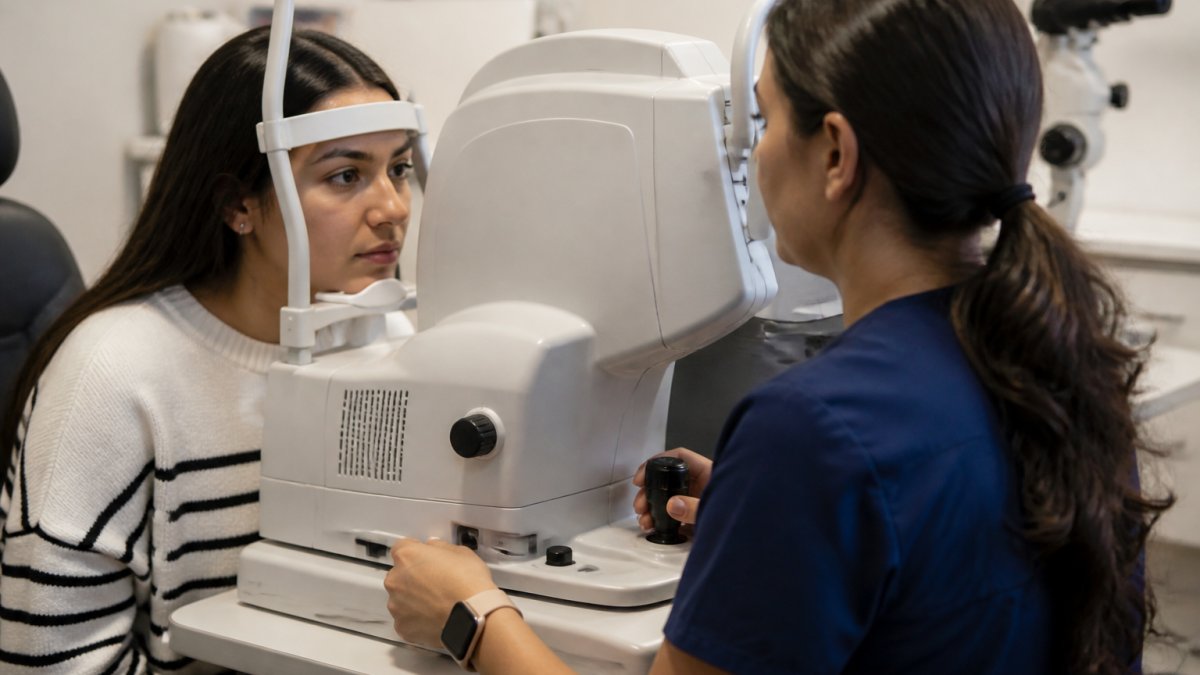
The scan itself takes seconds. You may see a brief flash of light but feel nothing as the machine captures multiple cross-sectional images of the retina and optic nerve.
Image quality depends on clarity and how still you keep your eye. If you move significantly, the scan may need repeating, and your technician will let you know.
After the scan, there are no restrictions. The images are available to your eye doctor immediately, either displayed on a screen in the exam room or reviewed later as part of your comprehensive eye evaluation. (Source: the American Academy of Ophthalmology’s guidelines on age-related macular degeneration)
Understanding Your OCT Scan Results and What They Mean
An OCT report can look intimidating, colored thickness maps, cross-sectional B-scans, and numbers compared against a reference range. Here is how to read the main elements and, just as importantly, how OCT fits alongside the other eye tests your doctor uses.
The main views on an OCT report
- Cross-sectional B-scan. A side view through the retina or optic nerve showing each layer. Fluid, drusen, and thinning show up here as changes in the normal layered pattern.
- Thickness map. A top-down color map of retinal or nerve fiber layer thickness. Warm colors (red, yellow) usually flag thinning or thickening outside the normal range; green is typically within normal limits.
- Reference-range comparison. Your measurements are compared to a database of age-matched normal eyes. A value in the 1st percentile is more concerning than one in the 20th percentile, even if both are technically ‘below average.’
- Change analysis. When you have prior scans, the software overlays them to show whether thickness is stable, improving, or progressing. This is often more informative than any single scan.
What the numbers do and do not tell you
OCT measures structure, the physical thickness and contour of retinal layers and the optic nerve, not function, or how well you actually see. A scan can show thinning before you notice vision loss, and it can also look stable in an eye that is functionally declining. That is why OCT findings are always read alongside your symptoms, visual acuity, and other tests.
How OCT compares to other eye tests
| Test | What it measures | What OCT adds |
|---|---|---|
| Dilated fundus exam | Direct view of the retina and optic nerve | Layer-by-layer thickness and fluid that may not be visible on exam |
| Retinal photography | A color image of the back of the eye | Cross-sectional detail and quantitative thickness measurements |
| Visual field testing | Peripheral and central visual function | Structural evidence that can precede functional loss |
| Tonometry | Intraocular pressure | Optic nerve and nerve fiber layer status, which pressure alone does not capture |
| Fluorescein angiography | Blood flow and leakage in retinal vessels | Non-invasive structural detail without dye injection |
These tests are complementary, not competing: OCT often detects a structural change first, visual field testing confirms a functional effect, and tonometry or angiography helps explain the cause. No single test tells the whole story.
Why a professional should interpret the report
OCT software flags values outside a reference range, but a flag is not a diagnosis. Anatomical variation, image quality, eye movement, and media opacity from cataracts can all shift the numbers, so an eye-care professional weighs the scan against your history, symptoms, and other findings. If a result looks abnormal, the next step is usually a repeat scan, an additional test, or a referral, not an immediate diagnosis.
Treat your OCT report as one chapter in your eye-health story. The scan shows structure; your symptoms and other tests show function. Together they give your doctor the full picture.
Who Should Get OCT Scans and How Often
OCT screening is most valuable for people whose eyes are at risk for conditions OCT can catch early. No single schedule fits everyone, but well-established risk categories guide who gets scanned and how often.
Risk-based guidance
People with diabetes. Diabetic retinopathy and diabetic macular edema can develop without symptoms. For type 1 diabetes, screening typically begins within five years of diagnosis; for type 2, at diagnosis.
People at risk for glaucoma. Risk factors include elevated intraocular pressure, family history of glaucoma, older age, African, Hispanic, or Asian ancestry, thin central corneas, and high myopia.
People at risk for age-related macular degeneration (AMD). Risk rises with age over 60, smoking, family history of AMD, high blood pressure, and obesity. A baseline macular OCT documents drusen and retinal thickness.
People with existing retinal disease. For patients already diagnosed with glaucoma, AMD, diabetic retinopathy, epiretinal membrane, or a macular hole, OCT becomes a routine monitoring tool.
What OCT is not a substitute for
A normal OCT does not rule out every eye disease. Conditions such as early cataracts, corneal disease, and some optic neuropathies are better assessed with other tools. OCT is one part of a comprehensive exam, not a stand-alone screening test.
Cost and coverage considerations
OCT is widely available in ophthalmology and optometry practices. When ordered to evaluate or monitor a diagnosed condition, glaucoma, AMD, diabetic retinopathy, or a suspicious finding, it is typically billed to medical insurance under the medical benefit, subject to deductible and coinsurance. As an optional add-on during a routine vision exam, it is often self-pay, with prices varying widely by practice and region.
Because coverage depends on the diagnosis code and reason for the scan, ask the practice two questions before your visit: (1) Is this billed to medical insurance or as an elective service? and (2) What is my out-of-pocket responsibility after my plan processes the claim? Practices can usually provide a good-faith estimate.
The bottom line
If you have diabetes, a family history of glaucoma or AMD, are over 50, or already have a retinal diagnosis, OCT is likely part of your care. The right interval is a clinical decision based on your risk profile, baseline scan, and how your eyes change over time. Ask your eye doctor what schedule fits your situation, and ask about cost and coverage at the same time so there are no surprises.
Conclusion
OCT imaging has transformed how eye doctors detect and monitor conditions that threaten vision. It reveals structural changes in the retina and optic nerve long before symptoms appear, creating an opportunity for early intervention when treatment is most effective. Whether you’re at risk for glaucoma, macular degeneration, diabetic retinopathy, or other retinal diseases, OCT scans give your doctor the detail needed to protect your vision.
If you’re over 50, have a family history of eye disease, or manage conditions like diabetes that affect eye health, ask your eye doctor about OCT imaging at your next exam. Clear Vision San Antonio offers advanced OCT technology as part of our comprehensive eye care services. Our experienced eye doctors use OCT findings alongside other diagnostic tools to give you the complete picture of your eye health and create a personalized plan to preserve your vision.
Frequently Asked Questions
What is an OCT scan and how does it differ from a regular dilated eye exam?
An OCT scan uses light waves to create cross-sectional images of your retina and optic nerve with high-resolution detail. Unlike a dilated eye exam, which allows your eye doctor to view the retina through a microscope, OCT imaging captures precise measurements of retinal thickness and structural layers. Both tests are valuable; OCT provides quantifiable data about subtle changes that may not be visible during a standard examination, making it especially useful for monitoring conditions like glaucoma and age-related macular degeneration.
Can an OCT scan detect glaucoma before I have any symptoms or vision loss?
Yes. OCT scans can measure the thickness of your optic nerve fiber layer and detect structural damage years before you notice vision changes. Glaucoma often progresses silently, causing irreversible optic nerve damage without early symptoms. By establishing a baseline OCT scan and comparing follow-up scans, your eye doctor can identify disease progression and start treatment to slow or prevent further vision loss, making OCT a powerful tool for early glaucoma detection.
How often should I have an OCT scan?
Screening frequency depends on your risk factors and eye health status. If you have glaucoma, diabetes, age-related macular degeneration, or a family history of eye disease, your eye doctor may recommend annual or more frequent OCT scans. Adults over 40 with risk factors benefit from baseline OCT imaging to establish a reference for future comparison. Your eye care professional will recommend a schedule tailored to your individual needs during a comprehensive eye examination.
Is an OCT scan painful or does it require any special preparation?
OCT scans are completely noninvasive and painless. No dilation drops, needles, or contact with your eye is necessary. The procedure typically takes just a few minutes. You sit at the imaging device, rest your chin on a support, and focus on a light while the scanner captures cross-sectional images. Most patients experience no discomfort, and there is no recovery time, you can resume normal activities immediately after.