Table of Contents
- Why Thin Corneas Make ICL Surgery a Game-Changing Option
- LASIK vs ICL for Thin Corneas: A Clear Comparison
- ICL Surgery Candidacy Requirements: Are You a Good Fit?
- What to Expect: The ICL Surgery Procedure Overview
- ICL Surgery Recovery Time: A Week-by-Week Post-Operative Lifestyle Guide
- Understanding the Full Cost of ICL Surgery for Thin Corneas
- Technology, Patient Outcomes, and Why Your Surgeon’s Credentials Matter
- Complimentary Consultation: Your First Step Toward Clear Vision
Last Updated: June 10, 2026
Patients with thin corneas often hear the same discouraging news: you’re not a candidate for LASIK. ICL surgery for thin corneas changed the equation entirely, opening a path to clear vision that simply didn’t exist for this population before. At Clear Vision San Antonio, we guide patients through exactly this situation every day. Below, we’ll walk through how the procedure works, who qualifies, what recovery looks like week by week, and what the full cost picture includes.
Here’s what most people get wrong: thin corneas aren’t a defect to work around. They’re simply a measurement that rules out one procedure while pointing directly toward a better one.
Why Thin Corneas Make ICL Surgery a effective Option
ICL surgery for thin corneas is a refractive surgery solution for patients whose corneal tissue is too thin to safely undergo LASIK or PRK. The Implantable Collamer Lens (ICL) is placed inside the eye, between the natural lens and the iris, without removing or reshaping any corneal tissue.
LASIK achieves vision correction by ablating corneal tissue with an excimer laser. The thinner your cornea, the less tissue is available for safe removal. Patients below a certain thickness threshold face elevated risk of ectasia, a progressive thinning and bulging of the cornea that can permanently compromise visual acuity. ICL sidesteps this risk entirely because the cornea is never touched.
The EVO ICL is FDA-approved and designed for patients with nearsightedness, farsightedness, and astigmatism. According to FDA device approval documentation for EVO ICL, the lens has demonstrated strong safety and efficacy data across clinical trials.
For patients with thin corneas, ICL surgery isn’t a compromise. It’s often the superior option, preserving corneal integrity while delivering visual outcomes that rival or exceed LASIK results.
The procedure also preserves the option for future cataract surgery, since the ICL can be removed if needed, something neither LASIK nor PRK can offer.
LASIK vs ICL for Thin Corneas: A Clear Comparison
The real difference between LASIK and ICL for thin corneas isn’t about which procedure is "better" in the abstract. It’s about which is safe and appropriate for your specific corneal anatomy.
| Feature | LASIK | EVO ICL | PRK | SMILE |
|---|---|---|---|---|
| Corneal tissue removed | Yes | No | Yes | Minimal |
| Suitable for thin corneas | No | Yes | Limited | Limited |
| Reversible | No | Yes | No | No |
| Dry eye risk | Moderate | Low | Moderate | Low |
| Recovery to functional vision | 24-48 hours | 24-48 hours | 5-7 days | 3-5 days |
| Treats high myopia | Limited | Yes | Yes | Limited |
How LASIK Reshapes the Cornea, and Why Thin Tissue Is a Problem
LASIK works by creating a thin flap in the corneal surface, then using an excimer laser to remove tissue from the underlying stroma. Higher prescriptions require more tissue removal. For thin-cornea patients, there simply isn’t enough tissue to remove safely. When residual stromal bed calculations fall below safe thresholds, LASIK is contraindicated, proceeding risks corneal ectasia, which can lead to severely distorted vision and may require corneal transplantation.
A common mistake is assuming "borderline" thin corneas are an acceptable risk. They aren’t. Experienced refractive surgeons don’t negotiate with that threshold.
How EVO ICL Works Without Removing Corneal Tissue
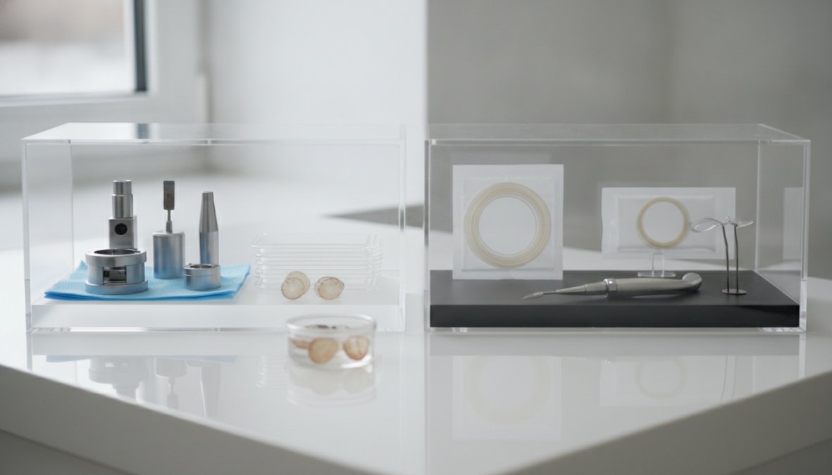
The EVO ICL is a collamer lens, roughly the size of a contact lens, folded and inserted through a small incision at the corneal edge. Once inside, it unfolds and positions itself in the posterior chamber, correcting refractive error by focusing light precisely onto the retina. No corneal tissue is removed, no flap is created, and the natural lens remains intact.
The EVO+ version includes a central port that allows aqueous humor to flow naturally, eliminating the pre-operative laser iridotomy that older ICL models required.
SMILE and PRK: Other Alternatives Worth Knowing
PRK doesn’t create a corneal flap but still removes corneal tissue via excimer laser, so it often remains contraindicated for genuinely thin corneas. SMILE removes less tissue than LASIK and may suit borderline-thin corneas with moderate prescriptions, but it is not a substitute for ICL when corneas are genuinely thin.
For patients with thin corneas and significant refractive error, ICL surgery typically offers the safest and most effective path to 20/20 vision.
ICL Surgery Candidacy Requirements: Are You a Good Fit?
The ideal ICL candidate is between 21 and 45 years of age, has stable vision for at least one year, and has adequate anterior chamber depth to accommodate the lens safely.
Key candidacy criteria include:
- Corneal thickness that rules out LASIK or PRK
- Anterior chamber depth of at least 2.8 mm
- Stable refractive prescription for a minimum of 12 months
- No history of certain autoimmune conditions or uncontrolled glaucoma
- Pupil size within acceptable parameters to minimize night glare
- Absence of significant cataracts
Patients outside the 21-45 age range are evaluated individually. Presbyopia patients should discuss realistic expectations, since ICL corrects distance and astigmatism but does not eliminate reading glasses in patients over 40.
Diagnostic Testing and Vision Stabilization Criteria
Vision stabilization is a hard requirement. A shifting prescription means the eye is still changing; implanting a fixed-power lens before stabilization leads to residual refractive error.
Diagnostic testing typically includes:
- Corneal topography: maps the corneal surface and identifies irregularities
- Pachymetry: measures corneal thickness at multiple points
- Anterior chamber depth measurement: confirms sufficient space for the lens
- Wavefront analysis: identifies higher-order aberrations
- Retinal evaluation: rules out peripheral retinal issues
According to American Academy of Ophthalmology refractive surgery guidelines, comprehensive pre-operative assessment is the single most important factor in achieving optimal surgical outcomes.
Patients who self-report stable vision without documented prescription history are not the same as patients with verified stabilization. Surgeons need actual prescription records spanning at least 12 months. Proceeding without this documentation affects long-term outcomes.
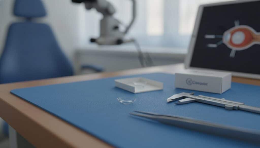
What to Expect: The ICL Surgery Procedure Overview
The ICL surgery procedure takes approximately 20-30 minutes per eye under topical anesthesia, eye drops, not injections. Patients remain awake but comfortable throughout.
The sequence:
- Dilating and numbing eye drops are administered
- A small incision (~3mm) is made at the corneal periphery
- The folded ICL is injected through the incision
- The lens unfolds and is positioned in the posterior chamber
- The incision self-seals without sutures in most cases
- The procedure is repeated for the second eye, often the same day or within a week
Most patients notice dramatically improved vision within hours. The absence of a corneal flap eliminates flap-related complication risk, and the small incision heals rapidly.
ICL Surgery Recovery Time: A Week-by-Week Post-Operative Lifestyle Guide
ICL surgery recovery is generally faster and more comfortable than PRK, and comparable to LASIK for functional vision restoration. Most patients return to desk work within one to two days. Understanding each phase prevents avoidable setbacks.

First 72 Hours: Restrictions and What to Watch For
Visual acuity improves rapidly, but the eye is still adjusting. Expect some light sensitivity, mild halos, and fluctuating clarity, normal effects that typically resolve within days.
Restrictions during the first 72 hours:
- No driving until cleared by your surgeon
- No eye rubbing under any circumstances
- Avoid extended screen use on the day of surgery
- Use prescribed antibiotic and anti-inflammatory drops on schedule
- Wear protective eye shields, especially during sleep
- No water contact with the eyes: no swimming, no hot tubs, no splashing
Set phone reminders for your eye drop schedule. Missing doses in the first week is the most common patient error and affects healing. Most surgeons prescribe drops on a tapering schedule over three to four weeks.
Weeks 1-4: Returning to Work, Driving, and Exercise
Most patients return to office work and driving within two to five days after the first follow-up confirms adequate visual acuity. Exercise follows a graduated timeline:
- Week 1: Walking only. No cardio, no gym
- Weeks 2-3: Light cardio and lower-body work permitted; avoid eye-impact risk
- Week 4: Most exercise resumes; contact sports require surgeon clearance
- After 4 weeks: Swimming typically cleared, with goggles required initially
Preservative-free artificial tears help with dry eye symptoms during extended screen use in this phase.
Understanding the Full Cost of ICL Surgery for Thin Corneas
ICL surgery typically costs more than LASIK, reflecting the higher cost of the lens implant itself. Cost varies by geographic market, surgeon experience, and technology used.
What drives cost variation:
- Surgeon experience and refractive fellowship credentials
- Diagnostic and surgical guidance technology
- Geographic location and practice overhead
- Whether both eyes are treated simultaneously or separately
Hidden and Often-Overlooked Costs Patients Should Budget For
The quoted procedure fee rarely represents the full financial picture. Costs that frequently catch patients off guard:
- Pre-operative diagnostic testing: workup fees may not be included in the procedure quote
- Post-operative medications: antibiotic and steroid drops add out-of-pocket cost without prescription coverage
- Follow-up visits: some practices include these; others bill separately
- Enhancement procedures: residual refractive error may require a touch-up (often PRK)
- Time off work: recovery restrictions may mean additional unpaid days for physical laborers
Ask your surgical coordinator for an itemized breakdown before committing. Total cost of care is what matters, not the headline price.
Financing and Payment Options That Make ICL Accessible
Most practices offer financing over 12-24 months, often with promotional interest-free periods. CareCredit and Alphaeon Credit are two widely accepted platforms covering elective vision procedures.
HSA and FSA funds can be applied to ICL surgery costs. According to IRS Publication 502 on medical and dental expenses, vision correction surgery qualifies as a tax-advantaged medical expense. Get the full itemized cost, calculate the HSA/FSA offset, then finance only the remainder, many patients find the net out-of-pocket cost more manageable than the sticker price suggests.
Technology, Patient Outcomes, and Why Your Surgeon’s Credentials Matter
Two factors drive outcome quality more than any other: the diagnostic technology used pre-operatively and the surgeon’s refractive surgery experience.
Advanced wavefront technology and corneal topography systems allow precise lens power selection, which directly determines whether a patient achieves 20/20 vision or retains residual refractive error. Board-certified ophthalmologists are the baseline, but refractive fellowship training indicates advanced specialization beyond general ophthalmology, higher ICL procedure volumes and deeper familiarity with edge cases like unusual anterior chamber geometries and high prescriptions.
Patients in their late 30s and 40s should also understand how the ICL interacts with eventual cataract surgery needs. A practice offering both services, like Clear Vision San Antonio, can plan across the full arc of a patient’s vision care rather than addressing each procedure in isolation.
As documented in peer-reviewed outcomes data on EVO ICL from the Journal of Refractive Surgery, patient satisfaction rates for ICL procedures among appropriately selected candidates are consistently high, with the majority achieving target visual acuity.
The surgeon’s diagnostic workup and refractive fellowship experience are more predictive of your outcome than any single piece of technology. Vetting credentials before choosing a provider is not optional.
Complimentary Consultation: Your First Step Toward Clear Vision
A complimentary consultation is where the abstract becomes concrete. No article or online calculator can tell you whether you’re a candidate for ICL surgery, only a comprehensive pre-operative evaluation can.
At a proper consultation, you should receive:
- Full corneal topography and pachymetry results
- A clear explanation of which procedures you qualify for and why
- An itemized cost breakdown with financing options
- Realistic expectations for visual outcomes based on your specific prescription
- A timeline for the pre-operative process and surgery scheduling
Patients who come to consultations informed ask better questions and make better decisions. This guide has given you that foundation. The next step is putting your specific anatomy and prescription into the equation.
For patients with thin corneas, the path to clear vision requires genuine expertise in ICL surgery, not just familiarity with it. Clear Vision San Antonio offers comprehensive pre-operative diagnostics, advanced technology lens implants, and personalized patient attention from initial evaluation through post-operative care. Request an appointment with Clear Vision San Antonio and take the first concrete step toward vision correction matched to your actual anatomy.
Frequently Asked Questions
Why are thin corneas a problem for LASIK?
LASIK uses an excimer laser to remove corneal tissue and reshape the surface, correcting nearsightedness, farsightedness, or astigmatism. If your cornea is too thin, removing tissue creates a structural risk, the remaining cornea may not be strong enough to maintain its shape under normal eye pressure, potentially leading to a condition called ectasia. This is why board-certified surgeons require a minimum corneal thickness before approving LASIK candidacy. ICL surgery sidesteps this issue entirely by adding a lens rather than removing tissue.
Is ICL surgery safe for patients with thin corneas?
Yes. ICL surgery for thin corneas is considered a well-established refractive surgery option precisely because it does not alter corneal tissue. The EVO ICL implantable collamer lens is placed behind the iris without any corneal removal, making corneal thickness essentially irrelevant to surgical safety. Patients with thin corneas who are otherwise healthy candidates, stable vision prescription, appropriate eye anatomy, and no contraindicated conditions, are generally excellent candidates for ICL. A complimentary consultation with diagnostic testing will confirm your specific eligibility.
What are the candidacy requirements for ICL surgery?
ICL surgery candidacy requirements typically include being between 21 and 45 years of age, having a stable vision prescription for at least one to two years (vision stabilization), and falling within a correctable range for nearsightedness, farsightedness, or astigmatism. The anterior chamber depth of the eye must be sufficient to safely house the implant. Patients should not have certain conditions like uncontrolled glaucoma or active eye disease. Comprehensive diagnostic testing during your consultation will assess all of these factors to determine whether ICL is right for you.
How long is the recovery time after ICL surgery?
ICL surgery recovery time is generally faster than many patients expect. Most people notice significantly improved visual acuity within 24 to 48 hours of the procedure. Driving is typically permitted within a few days once your surgeon confirms adequate vision. Strenuous exercise, swimming, and contact sports are usually restricted for two to four weeks. Post-operative care appointments are scheduled at one day, one week, and one month to monitor patient outcomes. Some patients experience mild night glare or halos in the first few weeks, which typically resolves as the eyes adjust.
What is the minimum corneal thickness for ICL, and how does it compare to LASIK requirements?
Unlike LASIK, ICL surgery for thin corneas does not have a strict minimum corneal thickness requirement because no tissue is removed during the procedure. LASIK generally requires a minimum pre-operative corneal thickness of around 500 microns, with a residual stromal bed of at least 250 microns post-surgery. ICL candidacy is instead evaluated based on anterior chamber depth, pupil size, and overall ocular anatomy. This makes ICL the preferred refractive surgery option for patients whose corneas are too thin to safely undergo LASIK or PRK.
This article was written using GrandRanker